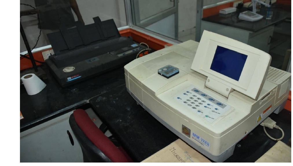
The UV-1800, a compact, double-beam UV-VIS spectrophotometer uses the Czerny-Turner mounting for its monochromator, and boasts the highest resolution in its class, a bright optical system, and a compact design. PC-controlled model, Shimadzu’s compact, double beam UV-1800 Spectrophotometer provides outstanding performance and functionality
A cell culture incubator is designed to maintain a constant temperature and high humidity for the growth of tissue culture cells under a CO2 atmosphere. Typical temperature settings range from 4C to 50C, and CO2 concentrations run from 0.3 to 19.9%. Non-corrosive stainless steel interiors are standard, but some newer models feature antimicrobial copper surfaces to prevent contamination. Auto decontamination using heat or UV light is another new and attractive feature available in CO2 incubators. Temperature in a CO2 incubator is typically controlled by a electric coils that give off radiant heat. Some units also include refrigeration for cooling. Relative humidity is maintained between 95% and 98% by an atomizer system or water reservoir. Features of cell culture incubators such as programmable controls with password protection, temperature alarms, CO2 alarms, and door opening alarms enhance convenience and security for the user.


BOD incubator is the most versatile and reliable low temperature incubator which is designed to maintain at 20°C, necessary for Biological Oxygen Demand/Biochemical Oxygen Demand (BOD) determination. BOD incubators provide controlled temperature conditions for accelerated tests and exposures. The biological oxygen demand (BOD) is an empirical test in which standardized laboratory procedures are used to determine the relative oxygen requirements of microbes in wastewaters, effluents, and polluted waters and in simple words, It is a chemical process that determines how fast biological organisms use up oxygen in a body of water or it measures the oxygen required for the biochemical degradation of organic material (carbonaceous demand) and the oxygen used to oxidize inorganic materials, such as sulfides and ferrous iron The seeding and dilution procedures provide an estimate of the BOD at pH 6.5 to 7.5.
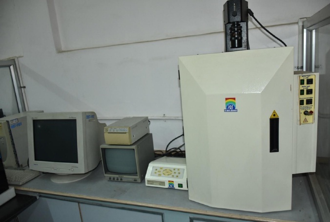
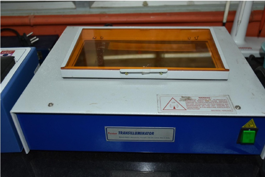
A gel doc, also known as a gel documentation system, gel image system or gel imager, refers to equipment widely used in molecular biology laboratories for the imaging and documentation of nucleic acid and protein suspended within polyacrylamide or agarose gels. These gels are typically stained with ethidium bromide or other fluorophores such as VisualaNA. Generally, a gel doc includes an ultraviolet (UV) light transilluminator, a hood or a darkroom to shield external light sources and protect the user from UV exposure, and a CCD or CMOS camera for image capturing.


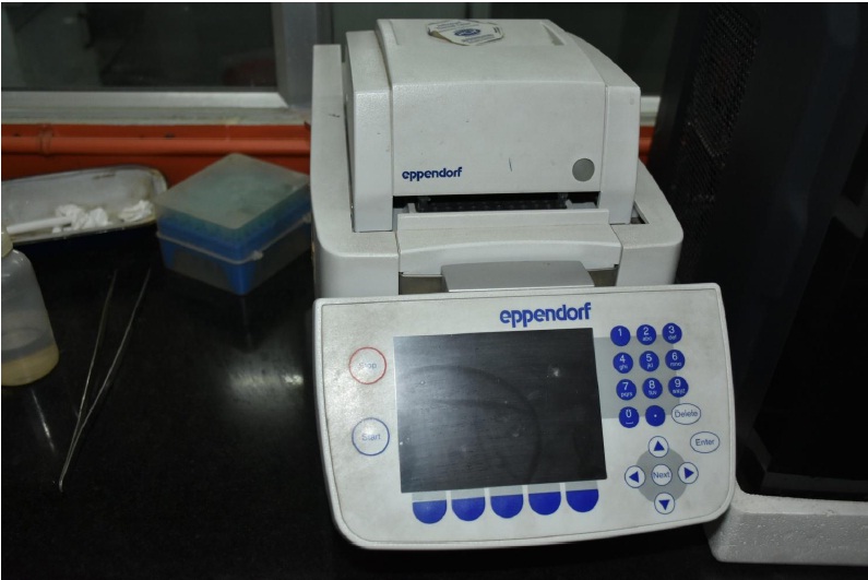
The ABI GeneAmp™ system 2700 is a simple thermal cycler with a small footprint, fit easily on the researcher’s personal work bench. The desktop footprint measures only 36 x 21 cm (14.2 x 8.3”) and it stands 22 cm (8.7”) above the desk. Because the air-flow for the cooling system is located at the back of the unit, several machines can be stored and operated side-by-side, with no space between, for efficient space usage. The polymerase chain reaction (PCR) technique is ubiquitous in laboratories and is used in applications such as DNA sequencing, cloning, library generations, mutagenesis, expression profiling, and more. PCR amplifies DNA by copying the nucleic acid strands exponentially. Thermal cyclers denature and anneal DNA strands during amplification and reagents such as enzymes, nucleotides and buffers to build the novel DNA.
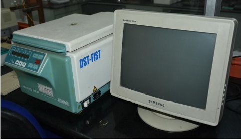
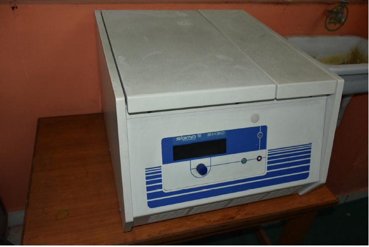
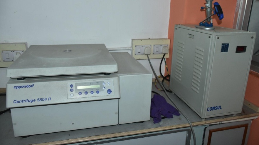
A centrifuge is a piece of equipment that puts an object in rotation around a fixed axis (spins it in a circle), applying a force perpendicular to the axis of spin (outward) that can be very strong. The centrifuge works using the sedimentation principle, where the centrifugal acceleration causes denser substances and particles to move outward in the radial direction. At the same time, objects that are less dense are displaced and move to the center. In a laboratory centrifuge that uses sample tubes, the radial acceleration causes denser particles to settle to the bottom of the tube, while low-density substances rise to the top. Very high speed centrifuges and ultracentrifuges able to provide very high accelerations can separate fine particles down to the nano-scale, and molecules of different masses.


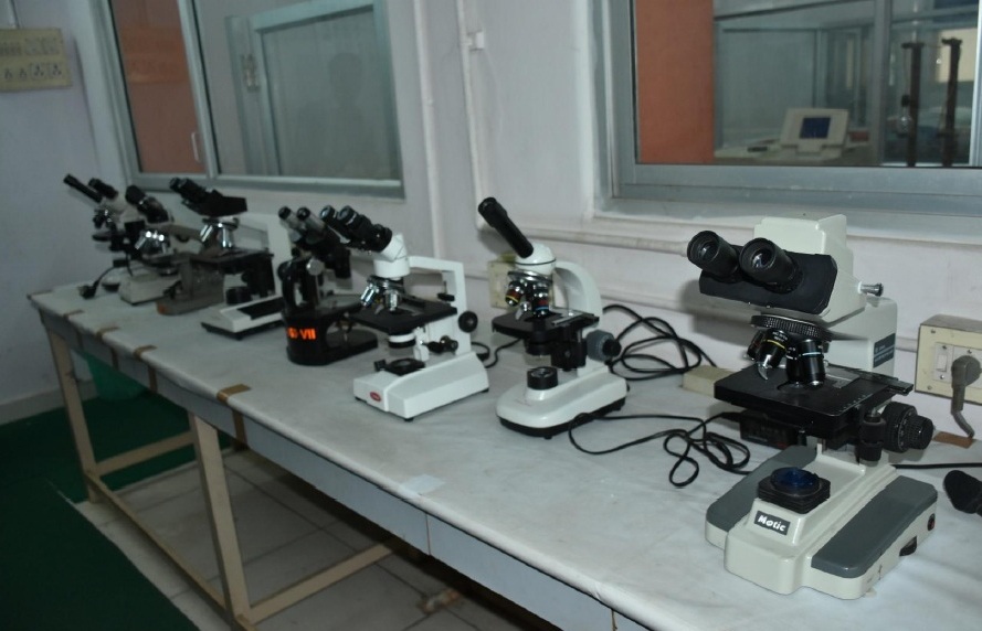
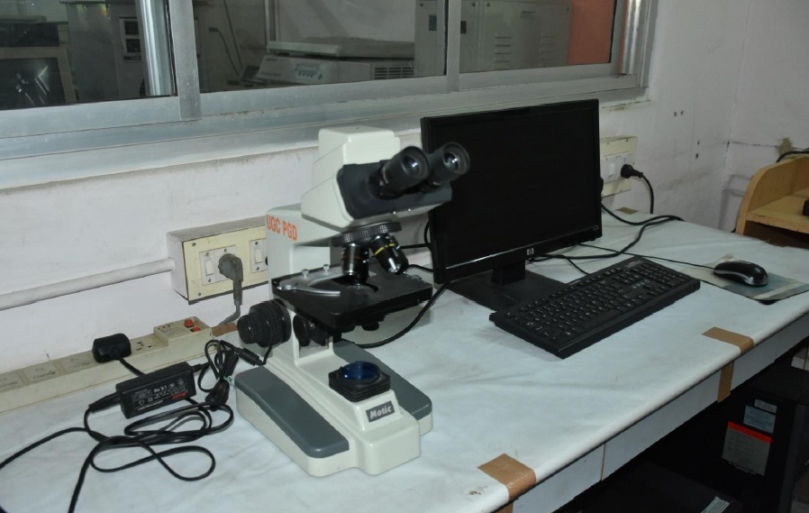
The stereo, stereoscopic or dissecting microscope is an optical microscope variant designed for low magnification observation of a sample, typically using light reflected from the surface of an object rather than transmitted through it. The instrument uses two separate optical paths with two objectives and eyepieces to provide slightly different viewing angles to the left and right eyes. This arrangement produces a three-dimensional visualization of the sample being examined.
Stereomicroscopy overlaps macrophotography for recording and examining solid samples with complex surface topography, where a three-dimensional view is needed for analyzing the detail.
The stereo microscope is often used to study the surfaces of solid specimens or to carry out close work such as dissection, microsurgery, watch-making, circuit board manufacture or inspection, and fracture surfaces as in fractography and forensic engineering. They are thus widely used in manufacturing industry for manufacture, inspection and quality control. Stereo microscopes are essential tools in entomology.
The stereo microscope should not be confused with a compound microscope equipped with double eyepieces and a binoviewer. In such a microscope, both eyes see the same image, with the two eyepieces serving to provide greater viewing comfort. However, the image in such a microscope is no different from that obtained with a single monocular eyepiece.
Some of the main strengths of Nikon microscopes include:
• Highest quality optics - Being the only company that manufacturers its own glass, Nikon strives to ensure the highest quality in production
• High durability of its mechanical components
• High precision
• Enhanced functionalities/capabilities to accommodate various research fields
• Impressive contrast levels, resolution and increased field of view
• A wide range of accessories


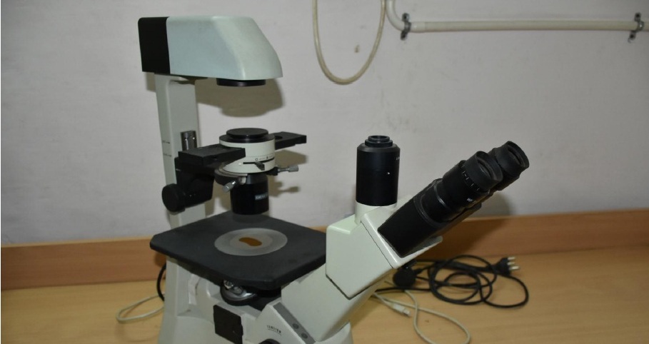
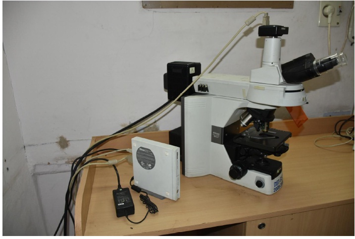
An inverted microscope is a microscope with its light source and condenser on the top, above the stage pointing down, while the objectives and turret are below the stage pointing up. The stage of an inverted microscope is usually fixed, and focus is adjusted by moving the objective lens along a vertical axis to bring it closer to or further from the specimen. The focus mechanism typically has a dual concentric knob for coarse and fine adjustment.
Depending on the size of the microscope, four to six objective lenses of different magnifications may be fitted to a rotating turret known as a nosepiece. These microscopes may also be fitted with accessories for fitting still and video cameras, fluorescence illumination, confocal scanning and many other applications.
Inverted microscopes are useful for observing living cells or organisms at the bottom of a large container (e.g., a tissue culture flask) under more natural conditions than on a glass slide, as is the case with a conventional microscope. Inverted microscope is also used for visualisation of the mycobacterium tuberculosis bacteria in the technique called microscopic observation drug susceptibility assay (MODS).
Phase-contrast microscopy is an optical microscopy technique that converts phase shifts in light passing through a transparent specimen to brightness changes in the image. Phase shifts are invisible, but become visible when shown as brightness variations. Changes in amplitude arise from the scattering and absorption of light, which is often wavelength-dependent and may give rise to colors.Phase-contrast reveals many cellular structures that are not visible with a simpler bright-field microscope, as exemplified in the figure. These structures were made visible to earlier microscopists by staining, but this required additional preparation and thus killing the cells. The phase-contrast microscope made it possible for biologists to study living cells and how they proliferate through cell division.


Used for storing biological samples for long duration at sub zero degree centigrade (< - 20°C) for biotechnological, microbiological and molecular studies.
A very common method for separating proteins by electrophoresis uses a discontinuous polyacrylamide gelas a support medium and sodium dodecyl sulfate (SDS) to denature the proteins. The method is called sodium dodecyl sulfate polyacrylamide gel electrophoresis (SDS-PAGE).


Southern blot analysis reveals information about DNA identity, size, and abundance. It is a classic technique that involves separating DNA fragments based on size via electrophoresis, transferring them to a membrane, hybridization with a labeled sequence-specific probe, washing, and finally detection of labeled DNA band(s). We offer one of the most comprehensive portfolios of products for Southern blot analysis.
The western blot (Protein immunoblot) is a widely used analytical technique in molecular biology, immunogenetics and other molecular biology disciplines to detect specific proteins in a sample of tissue homogenate or extract.


An autoclave is used to sterilize surgical equipment, laboratory instruments, pharmaceutical items, and other materials. It can sterilize solids, liquids, hollows, and instruments of various shapes and sizes. A very basic autoclave is similar to a pressure cooker; both use the power of steam to kill bacteria, spores and germs resistant to boiling water and powerful detergents.
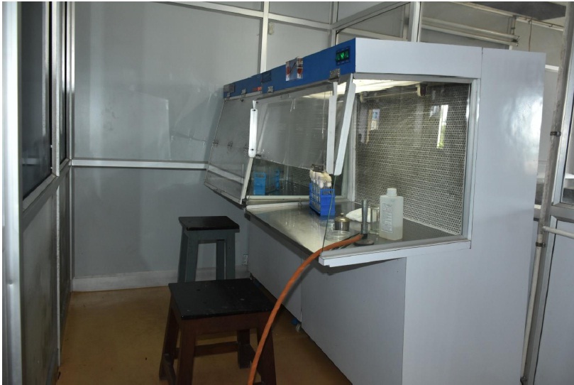
A laminar flow cabinet or tissue culture hood is a carefully enclosed bench designed to prevent contamination of semiconductor wafers, biological samples, or any particle sensitive materials. Air is drawn through a HEPA filter and blown in a very smooth, laminar flow towards the user. This instrument is useful for Microbiology and Animal Biotechnology practicals and UG, PG and Ph.D. research projects.


Provides controlled temperatures for doing specific temperature experiments. Any three specific temperatures can be set at a time and the experiments can be done simultaneously.
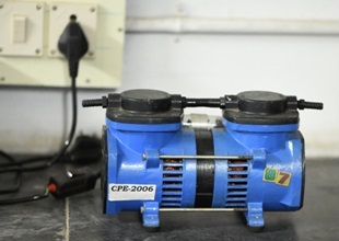
A vacuum pump is a device that removes gas molecules from a sealed volume in order to leave behind a partial vacuum. Vacuum pump mechanism is used to remove air or any gas particles from the container that creates vacuum in the vessel.

Specifications
Wavelength Range (nm): 335–1000
Wavelength Accuracy: ±2
Percent Transmittance Readability: 0.1%
Power Requirement: 110 V AC; 50 Hz
Dimensions: 16" W x 7" H x 12" D
These bottle shakers are use for mixing chemicals in Research Lab, Pharmaceutical Chemical/ Glass Plants, Bio Tech Lab, Educational Institute etc. for extraction of plant materials and for microbial culture and other preparations that needs constant stirring/mixing/shaking.